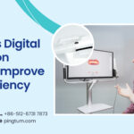
In traditional dentistry, X-rays are used to obtain the images of the teeth and gums, which are then studied by dentists to prescribe the treatment. Even though the quantum of X-rays used in such a procedure is minuscule, there is always a fear of radiation exposure. A 3D dental scanner has eliminated such risks and captures the images of the patient’s teeth and gums using a light source like a laser. In this procedure, the scanner projects light onto objects such as teeth, gums, dental arches, and implants, and converts the images into a 3D digital file called CAD files. Thereafter, the CAD files are sent to the dental laboratory through email where technicians create customized crowns, bridges, dentures, or other restorative solutions based on the recommendations of the dental surgeon and using AM technologies (Additive Manufacturing).
The best thing about these scanners is their ability to be quick and precise. They have the unique ability to capture multiple images of the buccal cavity and create a consolidated 3D model of the same. The benefits of any 3D dental scanner are as follows:
- Produces precise pictures of the mouth and teeth with dimensional accuracy
- Allows multiple images to be taken simultaneously
- Patients can view the real-time images simultaneously
- 3D models help patients to understand their dental issues better
- Allows simplification of treatment workflow by making it effective
- Ensures the safety and comfort of patients while scanning
- A higher rate of success
- A cost-effective process delivering better patient experiences
How does 3D scanning fare in dentistry
A digital scanner has plenty of usage for dentists, especially when it comes to making quick diagnoses and offering effective treatments.
Creating restorative solutions: Prosthetic appliances are in demand to treat patients with a broken or missing teeth. 3D scanners create an accurate model of the patients’ teeth and gums, which are then used to build prosthetic appliances. Making a best-fit model for such appliances can be a challenge, especially in traditional dentistry. However, in digital dentistry, the dental scanner can reproduce the exact dimensions of the patient’s buccal cavity. These help to construct the best-fit model for crowns, implants, bridges, and maxillofacial prostheses. For implants, the surgeon is guided by the model to drill at the precise location thereby saving time and assuring success.
Designing oral care objects: To foster better oral care among people, 3D dental scanners can help manufacture objects such as toothbrushes that are customized to the shape of the patients’ mouths. This new development can increase the convenience of patients while brushing and prevent the risks of inadequate oral care.
Surgical preview: The placement of prosthetic devices through surgery needs the dental surgeon to have a pre-surgical review and predict any issue. A 3D dental scanner can be used to reduce surgical risks and time.
Conclusion
As the world of dentistry evolves, the 3D dental scanner is going to carve a niche for itself even more. The quality output provided by such devices at high speeds has transformed both diagnosis and treatment and continue to deliver superior patient experiences.